


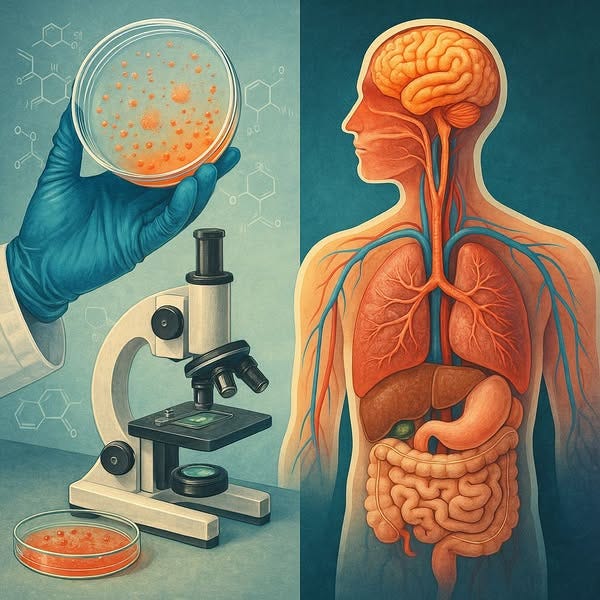
How does Electron Microscopy help sell the "virus" LIE?
Imagined "viral properties" are projected onto "particles" seen within Electron Microscopy images taken post the creation of a cytopathic effect through the use of a toxic brew, that's virology.

If you’re only going to look at one video linked in this article, this next one by Dr. Lee Merritt should be it. Otherwise this I will try to put together the best info exposing the Electron Microscopy part of the “Fear Thy Neighbour’s virus” fairytale.
The Flying Unicorn--Part 4: Electron Microscopy vs Light Microscopy and the Viral Theory (here)
Virologists can't "isolate" a supposed "virus"; they claim it’s a “dead” particle (code) outside the body, but comes alive (sequence) when inside the body. Virologists can't get an electron microscopy image when this hypothetical particle is allegedly outside the body, i.e, perhaps on a countertop. So, they claim a cytopathic effect of the sample taken from an ill patient, which they create through starving the cell culture sample, and then they introduce antibiotics, bovine serum, monkey kidney cells, trypsin, and other toxins.
Inventing the Nature of Viruses (here)
Tom Cowan asks why common-sense experiments have never been done with viruses. (here)
Indistinguishable Electron Microscopy images.
Which one’s the Spike Protein (here)
Appearances Can Be Deceiving - Viral-like Inclusions in COVID-19 Negative Renal Biopsies by Electron Microscopy (here)
The presence of identical "virus-like" particles in "uninfected" cell cultures shatters the illusion that these particles uniquely represent a "pathogenic virus." Their frequent presence where they shouldn't be should have raised serious questions amongst virologists, but didn’t.
Virus-like particles occur in cultures of stable pig kidney cell lines. Brief report (here)

An electron microscopy (EM) still image is captured, and a particle (debris, exosome) is “identified” by the virologist to be a “new virus.” Later, more of these “particles” are created for inclusion in the alleged “antibody-generating/immunizing” vaxsins. An adjuvant, i.e, Aluminum Hydroxide, also included in the vaxsin brew, is then used on a test subject to qualify that an antibody response has been stimulated, i.e, there is no “virus”/”specific antibody” determination (virology belief) required to qualify/obtain FDA “authorization”. “
…. An antibody does not react exclusively with the antigen that induces its appearance. Antibodies induced by and directed against a given protein may react with other proteins. Sometimes, many proteins”
“The scientific literature is replete with data showing antibodies are not very specific …” – Deni Papadopulos Eleopulos (Perth Group)
What's the story with "antibodies"? (here)
Most likely the so-called electron microscopy is just a technique by which particles of hard substances which are attached in an unverifiable way to fragments of organic matter, which are in natural decomposition and which are heavily damaged mechanically, chemically and thermally due to the preparatory laboratory procedures and due to the so-called X-rays, are micro-radiographed in a vacuum.
There is no possibility to confirm that the black and white shapes on the micrographs resulting from the radiography of particles of hard matter which are attached to particles of soft and transparent organic matter can provide uninterpretable evidence about the shape, size, external and internal structure and possible functions of the particles of residual organic matter which are impregnated with particles of hard matter.
No one knows even in the most superficial way what exactly exists and what exactly happens at the microscopic and especially at the submicroscopic level with the living organic matter that is extracted from the living being, that is detached from the vitality of the living being, that enters the natural process of decomposition, that is exposed to a new environment that is unnatural for it and that is damaged due to mechanical, chemical and thermal laboratory procedures.
Even more so, no one can know what exactly exists and what exactly happens at the microscopic and especially at the submicroscopic level with the living organic matter that exists in the living being.
To claim that by studying tissues that are dead and damaged using optical microscopy and so-called electron microscopy, we can find out what exactly exists and what exactly happens at the microscopic and especially at the submicroscopic level with the living organic matter that exists in the living being is the height of charlatanry.
Assumptions that are not confirmed by uninterpretable evidence are not science.
Humans do not have the sensory capacity nor the technological capacity to make direct and exhaustive observations in the microscopic realm, and especially in the sub-microscopic realm.
Humans do not know to what extent they can make direct and objective observations in the microscopic realm.
As for the submicroscopic realm, humans have no way of making even the most insignificant direct observation.
The fact that we can see different particles through optical microscopy is only a matter of little more value than nothing from the point of view of understanding what exists and happens in the microscopic realm.
Through optical microscopy, we cannot see particles in their original state, but particles that are a consequence of the mechanical, chemical, thermal, and energetic manipulation of the original matter from which they come.
The fact that we can see particles invisible to normal vision through optical microscopy does not mean that this is a guarantee that we can know exactly what exists and happens in the microscopic realm.
In reality, the human capacity to understand the functioning of existence is so limited that we can barely understand anything correctly, even in the realm visible to the naked eye.
This is also the reason why technology and medicine develop only on the basis of the method called trial and error, which involves carrying out an endless series of experiments done blindly and noting the effects in order to make technical applications based on them, following another endless series of experiments.
Apart from the statements related to the size, shape, and various peculiarities of the external structure of the particles seen through optical microscopy, all other statements made regarding the external and internal structural characteristics and functions of the said particles are pure speculation.
For example, no one has presented uninterpretable evidence that microscopic particles generically called germs possess the structural and functional characteristics required to be considered rudimentary forms of microscopic life.” - Gheorghe Constantin
Issues with Electron Microscopy Images claiming Viruses identification
“The electron microscope was invented in the 1930s, and they went looking in tissue samples, but they could not find anything different.” - Dr. Mark Bailey
Electron Microscopy can’t prove viruses exist (here)
Electron Microscopy and Unidentified “Viral” Objects (article-here) (video-here)
Thanks to Mike Stone of www.viroliegy.com for his efforts in unraveling the information below:
The Virus Concept (here)
Category: Purification-Isolation (here)
Category: Electron Microscope Images (here)
Category: Cell Cultures (here)
Helmut Ruska, often credited as the first person to image a "virus," admitted that the "newly discovered structures" observed in electron microscopy could have been artificial products created by the vacuum or electron beams. In other words, not "viruses." - Mike Stone
For my latest on the fraud of "virus-like" particles observed in electron microscopy:
Virus-like particles (here)
“Perhaps virologists use fetal bovine serum (FBS) in cell cultures to create "viruses" rather than human serum because “virus-like" particles are routinely found in FBS - even when labeled “virus-free" - whereas these same particles are absent in human serum.” – Mike Stone
Source:
Small, virus-like particles detected in bovine sera by electron microscopy (here)The Unethical Use of Fetal Bovine Serum (here)
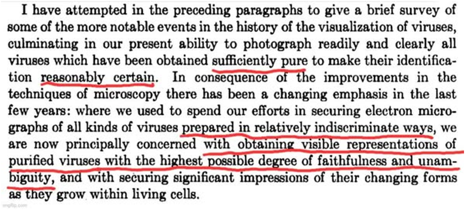
In 1954, Robley Williams noted that "virus" particles need to be purified (freed of contaminants, host materials, etc.) to the highest degree possible in order to draw a faithful and unambiguous conclusion they are the "virus." This was not the case for those already identified (Image 1).
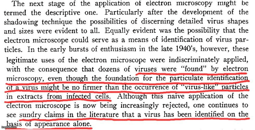
Williams discussed how "viruses" were identified in impure preparations. He stated that TMV could be identified in impure samples because of its rod-like shape, assuming all rod-like particles are the "virus." He noted EM could not identify unknown "viruses" in impure samples (Image 2).
Williams admitted that EM has technical limitations, making interpretations subjective. He noted the mental leap from grayscale images to assuming a "virus" is present and questioned whether they can ever be certain they're truly observing an "infectious" entity (Image 3).
Williams acknowledged that EM is often used on impure samples and cannot conclusively identify "virus" particles. He emphasized that EM can only show a correlation between a particle’s presence and an associated activity, but this does not demonstrate causation (Image 4).
Williams noted that "virus" identification requires more than images. A disease must first be established, & a distinct particle must be consistently correlated with it. He warned that EM’s visual appeal makes it vulnerable to misuse, leading researchers to see what they expect (Image 5).

Electron Microscopy of Viruses (here)
In 1956, Fawcett observed particles in frog tumors that resembled those claimed to be the herpes "virus" found in chick embryos by Morgan in 1954. He noted that "viral" nature is not established by images of particles claimed to be "viruses," & proof was still lacking.
Fawcett understood that just looking at particles in TEM & declaring they are "viruses" based upon previous "virus" images did not constitute proof.
Electron microscope observations on intracellular virus-like particles associated with the cells of the Lucké renal adenocarcinoma (here)


In 1957, virologist Robley Williams admitted that early "viruses" were "found" using electron microscopy without solid evidence that the particles were actually "viruses." He noted that numerous claims in the literature were made based solely on the appearance of the particles.
Source:
The Role of the Electron Microscope in Virus Research (here)
A 1964 paper reported that plants deliberately "infected" with an inoculant claimed to contain the tobacco mosaic "virus" (TMV) did not produce a single "viral" particle when examined using electron microscopy.
"Viral" particles were not detected at any stage, despite cytopathogenic effects occurring in the "infected" plants. Researchers suggested that CPE resulted from metabolic disruption, not "viral" replication, meaning the damage did not require a "virus" at all.


Source:
AN ELECTRON MICROSCOPE STUDY OF TOBACCO MOSAIC VIRUS LESIONS IN NICOTIANA GLUTINOSA L. (here)

The concern we now have is that EM results are assumed to represent “virus” qualification, and now the claim that genome sequencing is going to be the new way forward, all based on a false “isolation” foundation.
We reasoned that cryo-EM is mature for use in diagnostic virology (at genus level or finer) and pathogen discovery without the need for cultures or sequencing
Technologies for detection of uncultivatable viral pathogens by classical electron microscopy, 1, 2 serology, 3 or molecular biology techniques 4 have been largely supplanted by metagenomic or other sequence-based methods, 5 leading to a renaissance of diagnostic virology. 6 However, metagenomic approaches harbor pitfalls, including dependence on databases of known, annotated sequences and methodological biases.
Cryo-EM-based discovery of a pathogenic parvovirus causing epidemic mortality by black wasting disease in farmed beetles (here)
“If people were honest with themselves, they’d realize respiratory disease isn’t caused by invisible fictional boogeymen. The air we breathe is a major factor. No matter how many boosters you get, respiratory disease will persist as long as our air remains polluted.” – Mike Stone
Source:
Only seven countries met WHO air quality standards in 2024, data shows (here)Category: Air pollution (here)
Being from South Africa, I can’t monetize my substack.
If you’d like to buy me a SA cup of coffee, go - here,
For PayPal contributions, please go - here.
Thank you

https://linktr.ee/hewettinsit
https://andrew-hewett.pixels.com/